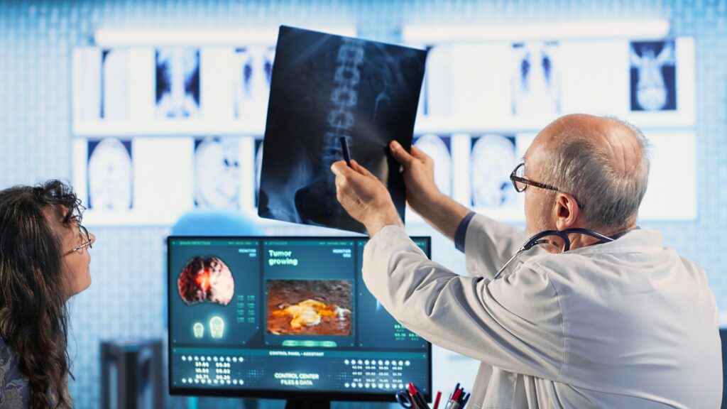
Skilled radiologist uses medical imaging technology to assess cancer risk factors, guiding old female patient through early detection and treatment options. Preventative medicine. Camera B.
A radiologist looks at hundreds of CT images to find a tiny shadow that could be cancer. At these moments, every pixel matters. AI can make that decision faster and more precisely today, but only if trained on perfectly labeled medical images.
Adding labels to diagnostic images isn’t just a technical step in AI research; it’s the most important one. Without labeled datasets, no medical AI model can be trusted to give consistent outcomes in real-world clinical settings. And in radiology, where the difference between “normal” and “abnormal” can be barely visible, that foundation is everything.
This blog explores the theoretical and practical gap in visual analysis by explaining the importance of diagnostic image annotation.
What is diagnosis image annotation?
Diagnosis image annotation involves labeling medical data from CT scans, MRIs, PET scans, ultrasounds, and X-rays so that computers can learn to spot specific patterns, problems, or conditions. AI models use these annotations, also known as “ground truth data,” to learn how to read images they haven’t seen before.
For instance, annotators might draw the exact edges of a tumor, mark where fluid is building up, or tag tissue that looks irregular. As the machine receives these annotated examples, it learns to identify features and recognize similar patterns in new cases. This process is similar to how a human radiologist becomes an expert by examining the patient’s medical scans repeatedly.
Why radiology needs precise annotated data
Given the success of ChatGPT, studies have been conducted on adapting it for downstream tasks. Researchers have investigated the quality of ChatGPT’s radiology report simplification and concluded that simplified reports were factually correct, complete, and not harmful to patients.
Radiologists examine thousands of images every week. There can be too much visual information, from brain scans to lung X-rays. It takes time. This is precisely why AI systems trained on annotated data have been developed to assist radiologists. Translating radiology reports into simple language with the help of AI models saves time, which can be utilized in other critical areas.
For instance, an AI tool trained to find early-stage lung nodules. If the system’s training data included well-annotated CT scans with expert-verified areas of interest, it could eventually detect similar nodules in new scans, even before a person does. This not only accelerates the diagnosis process, but it also makes it more accurate overall.
However, none of these improvements can happen without high-quality image annotation for radiology. The data that an AI algorithm learns from is what makes it work. If you put in insufficient data, you’ll get bad data out.
The contribution of medical experts in annotation
Radiology image annotation requires both technical and domain knowledge. Consider adding annotations to images to assist in identifying tumors. It’s not always easy to tell where the tumor starts and ends. It’s hard to set clear boundaries because of differences in tissue density, overlapping structures, and patients.
Board-certified radiologists review the training data for medical AI for clinical relevance and real-life applications. This process ensures datasets are both technically correct and diagnostically meaningful, enabling AI models to perform reliably in real-world medical settings.
Medical experts ensure that the data presented is more like what happens in the real world than just textbook examples. If an annotation is slightly faulty, it can confuse the AI model and lead to incorrect diagnostic results.
Challenges in image annotation
Despite several beneficial aspects of image annotation, it remains a challenging task that organizations must navigate.
- Volume of data: Medical imaging produces millions of unstructured data every day. Annotating even a small portion of this process is labor-intensive and time-consuming.
- Expert availability: Building an in-house team and hiring radiologists for image annotation is expensive. Due to their limited availability, they might even slow down workflows.
- Standardization issues: Different institutions often follow various annotation protocols, making it challenging to create universally reliable models. A domain-specific model requires
domain-specific training data, which is not fulfilled by synthetic data generated by AI tools.
- Tool limitations: While some annotation tools are robust, many are not optimized for complex tasks, leading to inefficiencies.
- Privacy: Protecting sensitive medical information is a top priority for both technical and ethical considerations, given that the images originate from real patient cases. Compliant training data is needed to ensure no hindrances when the model is ready for deployment.
Despite these challenges, investment in this space continues to grow, mainly because the potential returns in diagnostic accuracy and patient outcomes are too significant to ignore.
What the future holds
As AI continues to improve, the future of image diagnosis annotation will move toward semi-automated pipelines. These pipelines will use existing models to make first-pass annotations, which experts can review. Advanced radiology departments and research centers already use this human-in-the-loop method, which balances speed and accuracy.
Furthermore, the push for federated learning and privacy-preserving data methods could allow annotation to grow without compromising medical data privacy. These new ideas will not only accelerate the growth of AI in radiology but also enhance its accessibility for everyone around the world.
In the future, diagnostic image annotation will be part of a larger medical data system that is easy to read and can be used by different computer vision technologies.
Why does this matter?
The application to real-life-saving healthcare solutions, such as finding cancer early or spotting strokes with precise training data that models analyze, holds significant importance. This includes saving time and automating analysis for medical practitioners by identifying irregularities in reports faster that might have otherwise gone unnoticed.
Behind every correct diagnosis an AI system makes is a vast amount of annotated data from experts who know much about machines and medicine. In other words, diagnosis image annotation is the unseen driving force behind some of the most exciting new developments in medical AI. It isn’t showy, but it’s essential.
Conclusion
When people talk about healthcare AI, getting caught up in the end products, like smart assistants, diagnostic tools, and predictive algorithms, is easy. However, all of these systems depend on reliable data, and that data only makes sense when experts at anolytics accurately annotate it.
As AI becomes more common in radiology, image annotation for diagnosis will remain essential. It transforms images into information and medical scans into quantifiable actions.
The next time you hear about a medical AI breakthrough, remember that it all started with a human expert, a digital image, and a well-placed note.